
Gallery 24: Electron micrograph of RNA/DNA hybrid
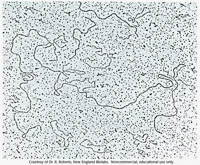
This was one of the original photos that Roberts and his group used for analyzing their results.
Electron micrograph, RNA, DNA, hybrid
- ID: 16533
- Source: DNALC.DNAFTB
Related Content

16541. Video 24: Richard Roberts, clip 5
Performing the electron micrograph DNA/RNA hybridizations, and seeing the results.

16636. Gallery 29: Electron micrograph of chromatin
Electron micrograph of the 10-nm fiber.

16637. Gallery 29: Electron micrograph of chromatin (1)
Electron micrograph of the 30-nm fiber.

16767. Gallery 37: Normal Drosophila Head, electron micrograph
Scanning electron micrograph of the head a normal Drosophila.

16638. Gallery 29: Chromosome with histone stripped
Electron micrograph of the DNA and the protein scaffold left over from one chromosome (insert) with all the histone stripped out.

16528. Concept 24: The RNA message is sometimes edited.
RNA splicing removes non-coding introns and splices together exons.

16649. Gallery 30: An electron micrograph of a mouse liver cell
An electron micrograph of a mouse liver cell. Magnification approximately 12,000 times.

16768. Gallery 37: Antennapedia Drosophila Head, electron micrograph
Scanning electron micrograph of the head a Drosophila mutant for the antennapedia gene.

16550. Problem 24: The RNA message is sometimes edited.
Map DNA molecules using restriction enzymes.

16529. Animation 24: The RNA message is sometimes edited.
Rich Roberts and Phil Sharp explain restriction enzymes, electrophoresis, and split genes.