
Gallery 25: Infection of H-9 cells with the MN strain of HIV-1 virus, electromicrograph 2
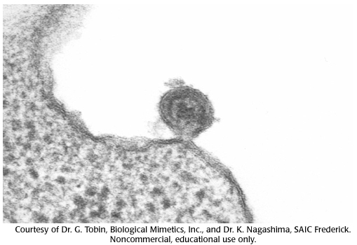
Virus particle is fusing with the cell membrane and about to empty its contents into the cell. Note the visible inner core.
virus particle, inner core, cell membrane, hiv, cells
- ID: 16559
- Source: DNALC.DNAFTB
Related Content

16560. Gallery 25: Infection of H-9 cells with the MN strain of HIV-1 virus, electromicrograph 3
Virus particle budding out from the cell. Although similar to the previous micrograph, the inner core is not as dense and therefore this is an "immature" viron budding out as opposed to a mature virus fusing in to infect a cell.

16558. Gallery 25: Infection of H-9 cells with the MN strain of HIV-1 virus, electromicrograph 1
Magnification including computer enhancements are approximately 100,000 times. A mature round virus particle is sitting next to the cell ready for infection. Note the visible inner core.

16561. Gallery 25: Infection of H-9 cells with the MN strain of HIV-1 virus, electromicrograph 4
Mature virus particles released from host cell.

16135. HIV jumps barrier zoonosis
Image showing Watson and Crick, chimp, macaques, HIV virus particles.

16552. Animation 25: Some viruses store genetic information in RNA.
David Baltimore and Howard Temin explain work on the Rous sarcoma virus.

16137. HIV Virion annotated
HIV particle proteins annotated.

1024. Pathways, Releasing the protein
In this section learn that newly made proteins leave the endoplasmic reticulum wrapped in a layer of membrane called a vesicle.

16087. Animal cell
Organelles in a typical animal cell.

16569. Problem 25: Some viruses store genetic information in RNA.
Explore the reverse transcriptase mechanism.

16877. Cell Signals
Journey inside a cell as you follow proteins and learn about cellular interactions. This 3-D animation brings to life the inner workings of a fibroblast cell as it responds to external signals. Created by Cold Spring Harbor Laboratory and Interactive Know